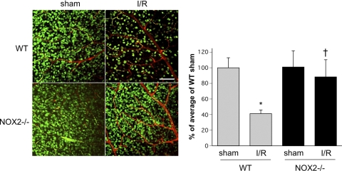
Figure 5.
Neuronal cell density in the GCL is preserved in the NOX2−/− retina during I/R. Confocal imaging of flatmounted retina labeled with NeuN antibody at 7 days after I/R shows a prominent decrease in density of NeuN-positive cells in the GCL of the WT I/R retina compared with the WT sham retina. NOX2 deletion significantly decreased the loss of NeuN-positive GCL neurons after I/R (n = 4; *P < 0.05 vs. sham; †P < 0.05 vs. WT I/R). Scale bar, 100 μm.

