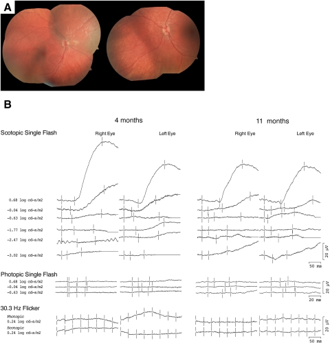
Figure 1.
(A) Fundus photographs from the case 1 patient at 4 months of age demonstrating a normal-appearing fundus. (B) Full-field ERGs from the case 1 patient at 4 and 11 months of age. Photopic ERGs were unrecordable at both time points. Scotopic ERGs were unrecordable at dim intensities, but at higher intensities a slow positive waveform was elicited.

