Figure 2.
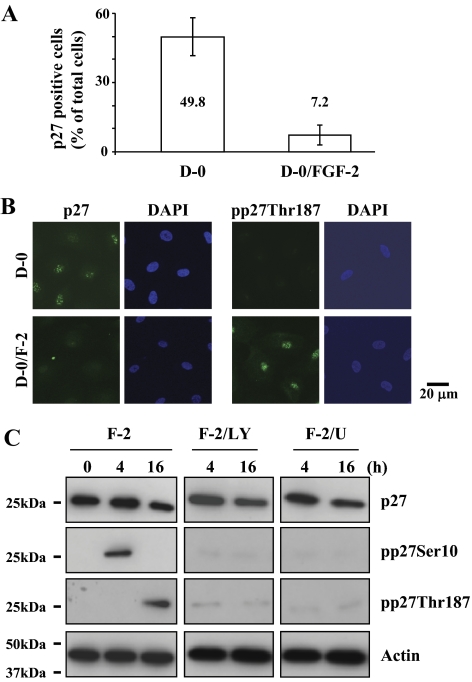
Reduction of p27 level ex vivo and induction of its phosphorylation at both Ser10 and Thr187 sites by FGF-2 stimulation in vitro. (A) The corneal endothelium was incubated with or without FGF-2 for 24 hours and then immunostained with anti-p27 antibody. The total DAPI-stained and p27-positive cells were counted in one microscopic field under high magnification (×400). The percentage of p27-positive CECs was significantly greater in the corneal pieces incubated in mitogen-deprived medium (D-0) than in those treated with FGF-2. (B) The serum-starved hCECs were treated with or without FGF-2 for 16 hours and maintained in D-0 for up to 24 hours. The cultured cells were fixed and labeled with anti-p27 or anti-pp27Thr187 antibody (FITC) and DAPI, respectively. (C) The serum-starved hCECs were pretreated with PI 3-kinase inhibitor or MEK1/2 inhibitor for 2 hours and then maintained in DMEM with FGF-2 for the designated time. At the end of treatment, cells were lysed and then immunoblotted with the respective antibody. Actin was used to control protein concentration on immunoblot analysis. The results represent data obtained in three independent experiments.

