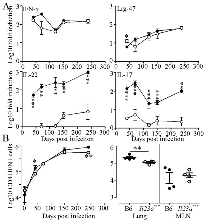
Figure 2. Mice lacking IL-23 maintain expression of IFN-γ and LRG-47 but not IL-17 or IL-22.

(A) B6 (filled circles) and Il23a−/− (open circles) were infected as for Fig 1 and induction of mRNA for IFN-γ, Lrg-47, IL-17 and IL-22 relative to uninfected control mice determined by RT-PCR. One of two representative experiments is shown. (B) A single cell suspension from infected mice was analyzed by flow cytometry (left panel) or ELISpot (right panel, Day 238) for IFN-γ production. Data points are the mean ±standard deviation for an n of 3–4 mice per group. Significance was determined by the Student’s t test with * P ≤ 0.05, ** P ≤ 0.01, *** P ≤ 0.001.
