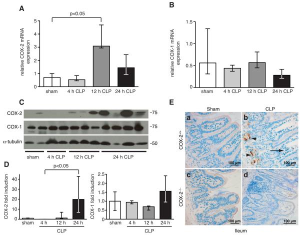
FIGURE 1.
COX-2, but not COX-1, is upregulated in the ileum following peritonitis-induced polymicrobial sepsis. RNA was isolated from ileums of COX-2+/+ mice following sham or CLP (n=2-3), reverse-transcribed, and qPCR performed for COX-2 (A) and COX-1 (B). Results were normalized to endogenous control 18S (p<0.05 by the Mann-Whitney test). (C) Protein was harvested from ileums of COX-2+/+ mice (n=2-4) following sham or CLP and Western blot performed for COX-2 and COX-1. (D) Bar graphs represent protein levels (normalized for α-tubulin) as determined by densitometry. Protein levels are expressed as fold induction compared with COX-2+/+ mice following sham (p<0.05 by the Kruskal-Wallis one-way ANOVA test followed by Dunn’s post test). (E) Representative COX-2 immunostaining of ileums harvested from COX-2+/+ and COX-2−/− mice 24 h following sham or CLP. Arrows represent positively stained inflammatory cells in the lamina propria and arrowheads represent positively stained epithelial cells in the ileum.

