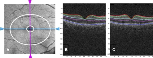
Figure 1.
Cirrus OCT en face image (A) displaying the 6 × 6 mm portion of the retina scanned by the acquisition protocol with the annulus (area between the two white rings) within the cube used by the Cirrus GCA algorithm to measure the thickness of the GCIPL. (B, C) Segmentation of macular intraretinal layers from a horizontal and a vertical tomogram, respectively, with the Cirrus GCA algorithm. Boundaries (top to bottom): red, internal limiting membrane; green, RNFL-RGC boundary; light blue, IPL-INL boundary; magenta, IPL-OPL boundary; dark blue, Bruch's membrane. Layers (top to bottom): RNFL, GCIPL, internal nuclear layer (INL), and OPL/photoreceptors.

