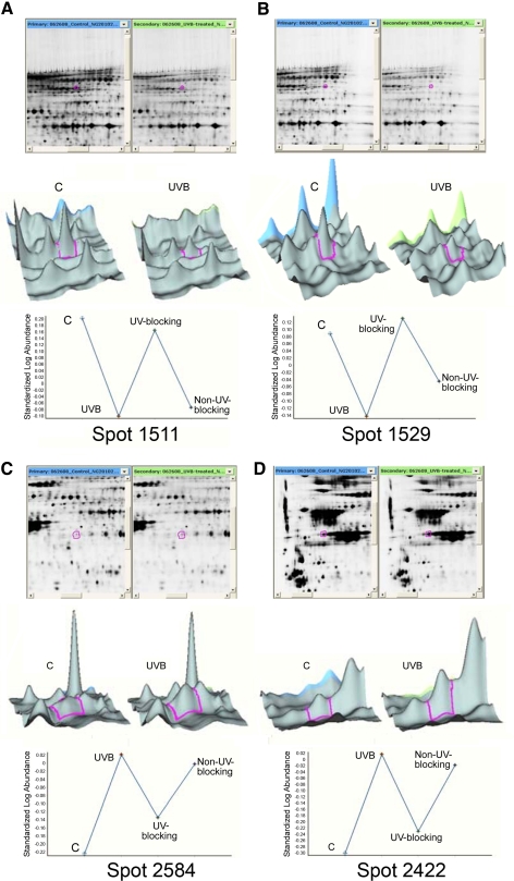
Figure 3.
Analysis of changes in lens epithelial cell proteins induced by UVB exposure in the absence of contact lenses and in the presence of UV-blocking and non–UV-blocking contact lenses. HLE B-3 cells were exposed to 768 mJ/cm2 UVB radiation at 302 nm and then incubated in culture medium for 4 hours. Proteins were separated by 2D-DIGE and subjected to MS/MS analysis. Quantitative image analysis data for identified proteins are shown in Table 3. Top: image of the gel with the protein spot encircled (red); middle: 3D data representation; bottom: quantitation of the proteins contained within the spot. (A, B) Spots 1511 and 1529 represent two of the protein spots that decreased with UVB irradiation. (C, D) Spots 2584 and 2422 represent two of the protein spots that increased with UVB irradiation.

