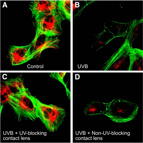
Figure 4.
Confocal microscopic analysis of F-actin and β-tubulin staining in HLE B-3 cells exposed to UVB radiation. HLE B-3 cells were exposed to 768 mJ/cm2 of UVB radiation at 302 nm and then incubated in culture medium for 4 hours. Cells were then fixed, stained with fluorescein phalloidin to visualize actin (green), and immunostained with an antibody to β-tubulin and Alexa 568–labeled secondary antibody (red). Note that UVB exposure reduced the F-actin and β-tubulin signals (B) compared with controls with no UVB exposure (A). This decrease was prevented by senofilcon A class 1 UV-blocking contact lenses (C) but not by non–UV-blocking lotrafilcon A contact lenses (D).

