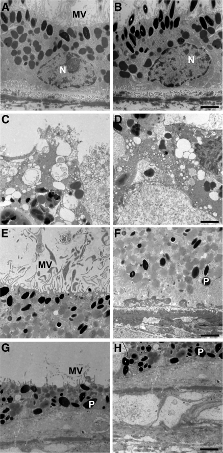
Figure 12.
RPE degeneration in an RPE65 postmortem donor eye. Electron micrographs of the RPE from the control (A, B) and the RPE65 donor (C–H). The control RPE displayed apical microvilli (A) and basal infoldings (B). The ultrastructure of the RPE65 donor RPE showed degenerating changes in the different quadrants studied. In the macula, apical microvilli were absent, and pleomorphic inclusions were common (C). The basal surface was characterized by the absence of infoldings and pleomorphic inclusions (D). In the nasal quadrant, apical microvilli were present (E). However, the basal surface was characterized by the presence of electron-dense material beneath the RPE cells (F). In the inferior quadrant, the RPE was discontinuous, and in some areas short apical microvilli were still present above the RPE (G) and a debris zone was present below it (H). N, nucleus; P, pigment; MV, microvilli. Scale bars, 2 μm.

