Figure 2.
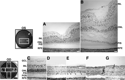
Degeneration in the retina of an RPE65 donor. Fundus images of both eyes with disc and macula delineated; schematic drawing of the regions cut and processed for cryosectioning (far left, top and bottom). One-micrometer plastic sections of both matched control (A, C) and RPE65 donor (B, D–G) postmortem retinas stained with toluidine blue. The macula of the left eye of the RPE65 donor (B) displayed edema. It contained a prominent preretina (epiretinal) membrane composed of fibroblastlike cells that were vitread to a connective tissue lamina (*). In the periphery, the retina of the RPE65 donor (D–G) displayed different degrees of retinal degeneration in all quadrants observed. Sparse inner and outer nuclear layers with stunted photoreceptor inner and outer segments are evident in the superior quadrant (E, arrowheads). A few pigmented cells are seen invading the degenerating retina of the temporal (F) and nasal quadrant (G, arrows). Finally, a thin, continuous area of pigmented RPE cells was present. Quadrants: M, macula; I, inferior; S, superior; T, temporal; N, nasal. RPE, retinal pigment epithelium; POS, photoreceptor outer segment; PIS, photoreceptor inner segment; ONL, outer nuclear layer; INL, inner nuclear layer; GCL, ganglion cell layer. Scale bars: 0.5 cm (fundus images upper and lower left); 400 μm (A, B); 200 μm (C–G).

