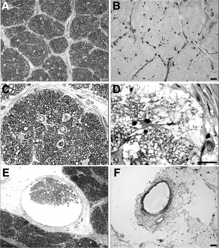
Figure 3.
Degeneration in the optic nerve of the RPE65 donor. Light microscopy of 1-μm plastic sections of optic nerve from both a matched control (A, C, E) and the RPE65 donor (B, D, F) stained with toluidine blue. Analysis of the optic nerve of the RPE65 donor in both low (B) and high (D) magnification showed the absence of myelinated axons when compared with the control (A, C). Only a few nuclei (astrocytes?) are evident (D). There is a paucity of axons consistent with the loss of ganglion cells in the retina. Finally, the RPE65 donor displayed a severely reduced central retinal vessel diameter (F). Scale bars, 200 μm.

