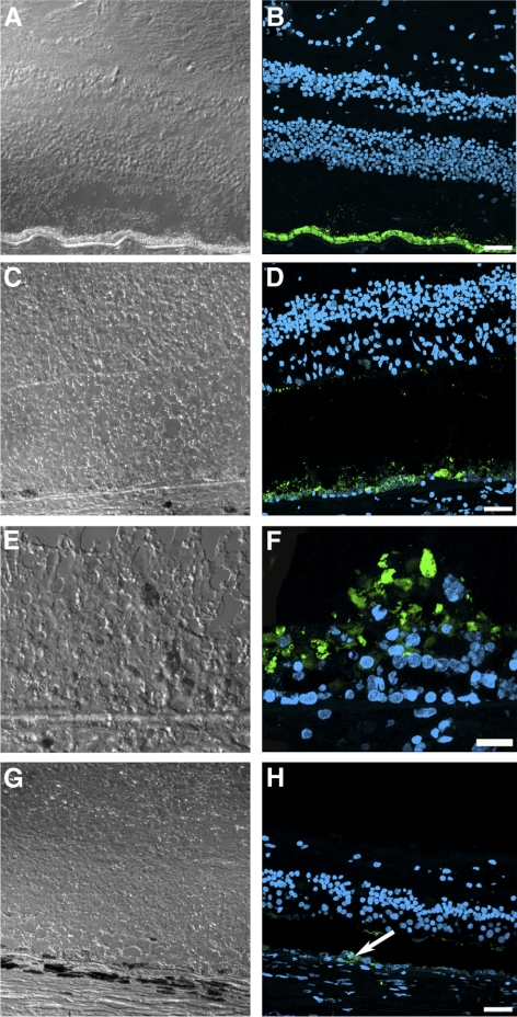
Figure 4.
Presence of remaining RPE cells expressing RPE65 in the postmortem eyes from an RPE65 donor. A continuous layer of cells was observed expressing RPE65 in the control (B) and in the macula of the RPE65 donor (D); however, the RPE65 donor expression was substantially lower than that observed in the control tissue. In a few areas in the macula of the RPE65 donor (F), several layers of RPE65-positive cells were observed in the RPE65 donor retina. In the periphery of the RPE65 donor retina, very few cells were detected expressing RPE65 (H, arrow). A, C, E, and G are differential interference contrast microscopy images of the same field shown in B, D, F, and H. Scale bars, 40 μm (B, D, H); 20 μm (F).

