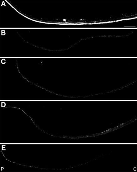
Figure 6.
Significant reduction in the cones in the periphery of an RPE65 postmortem donor eye. Montages of photomicrographs of the periphery tissue from the RPE65 donor (B–E) and control eyes (A) were analyzed using a cone arrestin antibody (7G6). Comparison of the samples showed that cones were mostly absent in the affected retina in inferior (B), superior (C), temporal (D), and nasal (E) regions. P, periphery; C, central. Scale bar, 500 μm.

