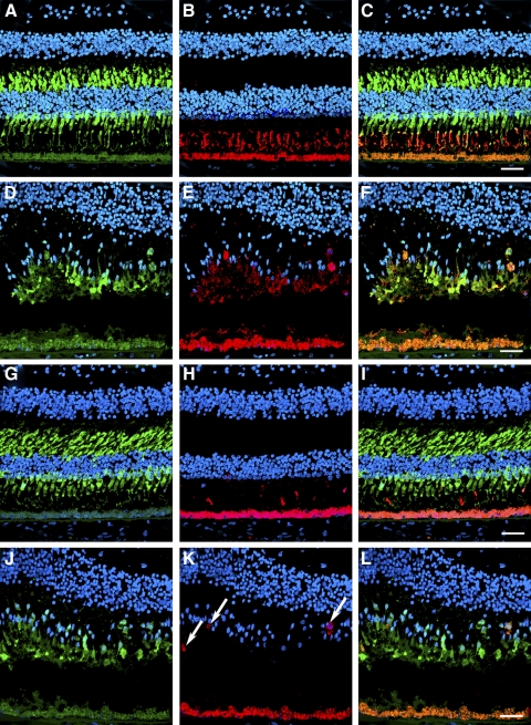
Figure 8.
Disorganized expression of red/green and blue opsins in the cones in the macula of an RPE65 postmortem donor eye. The distribution of cones was also analyzed in control and RPE65 eyes labeled with the cone arrestin (7G6), red/green opsin (AB5405), and blue opsin (AB5407) antibodies. Control retinas displayed cone arrestin distributed along the entire cone cell body (A, G), however the RPE65 donor retina displayed disorganized cones (D, J). In the control eyes, red/green (B) and blue (H) opsins were restricted to the cone outer segments. In the RPE65 eyes, the red/green opsin displayed a more diffuse staining (E) that overlapped with cone arrestin. However, blue opsin localization was mostly to the cone cell boundaries (K, arrows). Overlaid images are shown in C, F, I, L. Scale bar, 40 μm.

