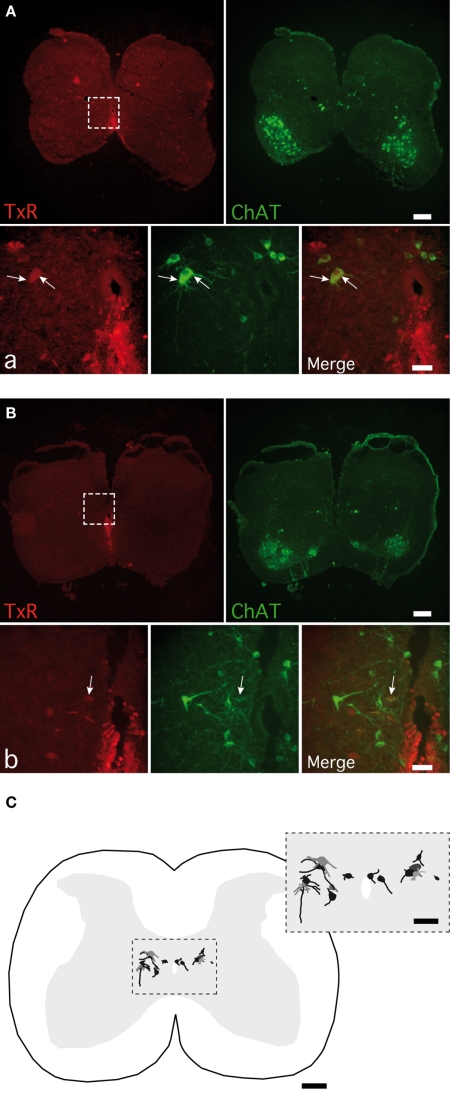
Figure 9.
Detection of cholinergic commissural interneurons in spinal cord slices. Photomicrographs of commissural interneurons (CINs) retrogradely labeled with Texas red (TxR), applied ventrally to the central canal and immunopositive for CholineAcetylTransferase (ChAT) in lamina VII (A) and close to the central canal (B). The areas boxed in (A,B) are shown at a higher magnification in (a,b) photomicrographs. Arrows point to the double-labeled CINs. (C) A normalized spinal cord transverse section illustrating the location of all the double-labeled CINs observed. The area boxed is shown at a higher magnification in the inset. [Calibration bars: (A–C), 200 μm; (a,b), 50 μm; inset, 100 μm].

