
Figure 1. Coupling pattern and dendritic stratification of OFF α-GCs in the rabbit retina.
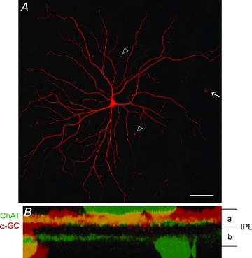
A, photomicrograph showing the flatmount image of an OFF α-GC injected with Neurobiotin. The somata of tracer-coupled amacrine cells (arrowheads) and α-GCs (arrow) can be visualized. Scale bar: 100 μm. B, a z-stack (vertical) confocal image of the dendrites of a Neurobiotin-injected OFF α-GC (red). The dendrites stratify within sublamina-a of the IPL. The two ChAT bands (green) were labelled using an antibody to provide landmarks for the boundaries of sublamina-a and -b.