
Figure 8. OFF α-GCs are coupled to amacrine cells that stratify within both sublamina-a and sublamina-b of the IPL.
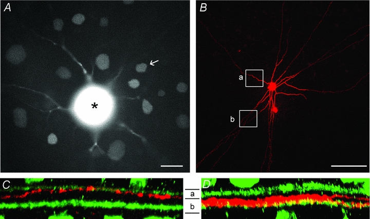
A, photomicrograph of the living rabbit retina showing an OFF α-GC injected with the gap junction-permeant fluorescent tracer Po-Pro-1. The soma of the OFF α-GC with bloomed fluorescence is indicated by the asterisk. The somata of coupled amacrine cell and ganglion cell neighbours can also be clearly visualized. One amacrine cell (arrow) was labelled with Neurobiotin. Scale bar = 25 μm. B, confocal micrograph of the amacrine cell in panel A labelled with Neurobiotin after post hoc histological processing. This subtype of amacrine cell typically showed long radiate branches that became narrow and axon-like, running for up to 2 mm. The rectangles show the portion of the processes in the confocal images in the subsequent panels. Scale bar = 100 μm. C, Z-stack vertical confocal images of the amacrine cell process (red) in rectangle ‘a’ in panel B showing that it stratifies in sublamina-a of the IPL. ChAT bands are shown in green. D, Z-stack vertical confocal image of the amacrine cell process (red) in rectangle ‘b’ in panel B showing that it stratifies within sublamina-b of the IPL.