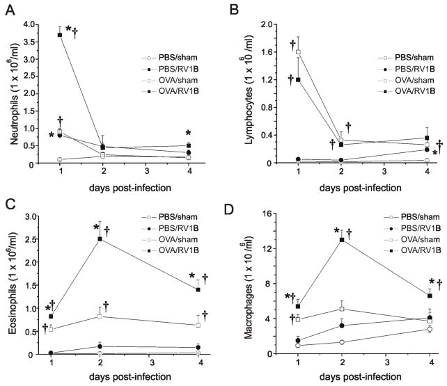
Figure 1. OVA/RV treated mice show increased tissue eosinophils and macrophages in response to RV infection.
Wild type BALB/c mice were sensitized intraperitoneally with endotoxin-free OVA and alum on days 1 and 7, and challenged intranasally on days 14, 15, and 16 with OVA. Controls were treated with PBS. Mice were inoculated with RV1B or sham (HeLa cell supernatant) on day 16. Mouse lungs were harvested 1, 2 and 4 after infection. Lungs were digested for 1 h in Type IV collagenase in serum free RPMI. Strained cells were treated with RBC lysis buffer, spun and enriched for leukocytes with 40% Percoll. Resulting pellets were resuspended in PBS and total cell count determined. Cytospins of leukocytes were stained with Diff-Quik and differential cell count determined for 200 cells. Time courses for tissue neutrophils (A), lymphocytes (B), eosinophils (C) and macrophages (D) are shown. (N=4-5 mice per group, bars represent mean±SEM, *different from respective sham group, †different from respective PBS group, P<0.05, one-way ANOVA.)

