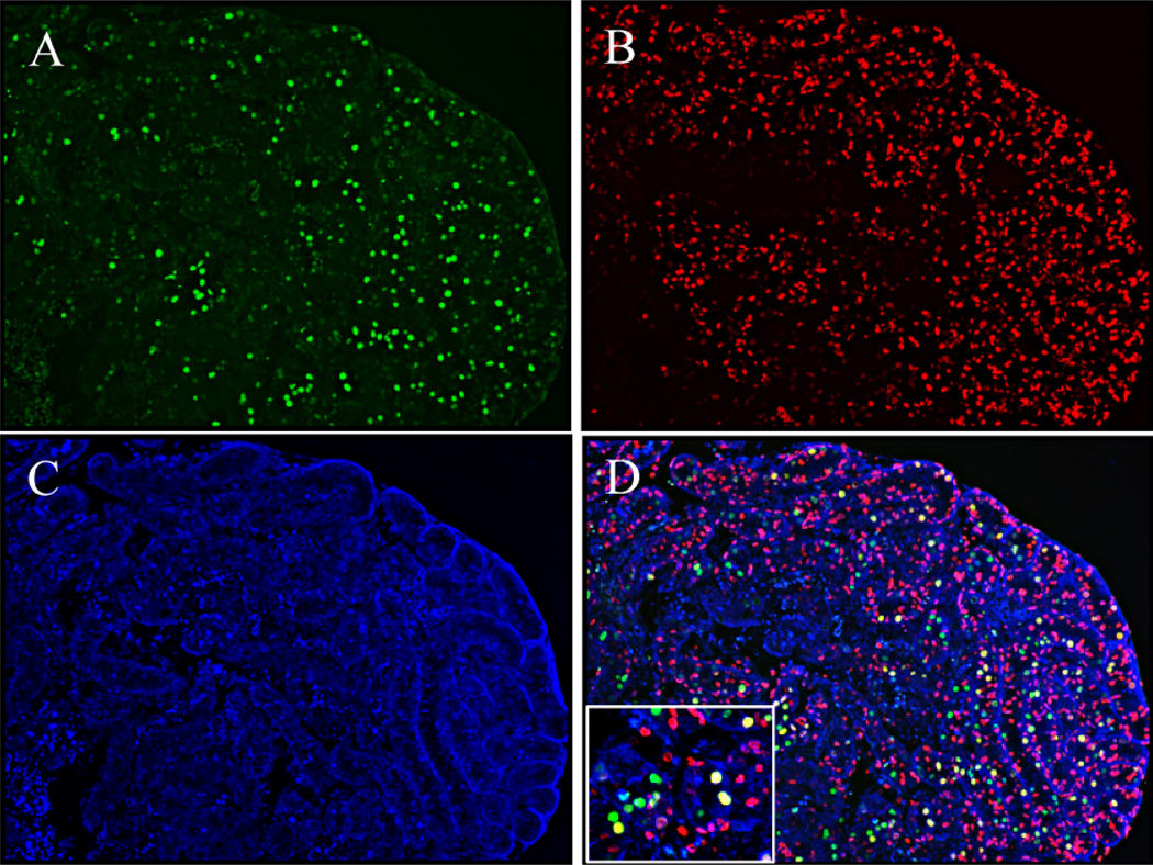
FIG. 2.
Immunohistochemical double staining of GCNA1 and BrdU in cultured neonatal testes. A) Immunostaining of GCNA1 (green). B) Immunostaining of BrdU (red). C) 4′,6-diamidino-2-phenylindole counterstaining of cell nuclei (blue). D) Overlay of images A, B, and C. Nuclei of proliferating germ cells appear yellow. Original magnification ×100 (A–D) and ×200 (inset).

