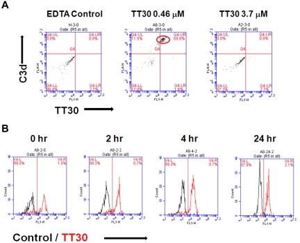
Figure 4.
Detection of TT30 on C3d-decorated rabbit RBC after hemolysis induced by CAP activation in human serum. Residual RBC protected from hemolysis were stained with biotinylated anti-C3d (A702; Quidel) and FITC-conjugated anti-CR2 (clone HB5; Santa Cruz Biotechnology) monoclonal antibodies to detect C3d fragment deposition and TT30, respectively. Streptavidin-conjugated APC (BD Biosciences) was used as a secondary detection reagent. Isotype controls were from BD Biosciences. Cells were analyzed on Accuri C6 cytometer (Accuri Cytometers Inc) using CFlow software. (A) TT30 bound to rabbit RBC protected from hemolysis at 0.46μM can be detected by flow cytometry (middle panel) in contrast to a higher bound TT30 concentration of 3.7μM (right panel). In presence of EDTA, there is no C3 fragment decoration of rabbit RBC and thus no binding of TT30 (EDTA control, left panel). Results represent dot plots of double staining of one representative experiment. (B) TT30 binding is retained on the RBC surface for at least 24 hours. Histograms represent overlays of 1-color staining of isotype control (black) over TT30 staining (red).

