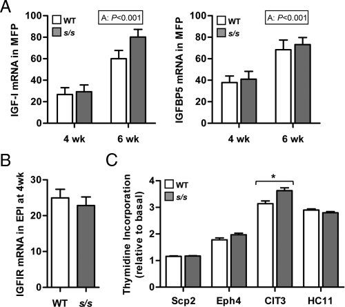
Fig. 3.
Effect of lack of leptin-dependent STAT3 signaling on the intramammary IGF-I system and on the ability of serum to stimulate mammary epithelial cell proliferation. A, MFPs were collected from female mice lacking leptin-dependent STAT3 signaling (s/s) or their WT counterparts at 4 and 6 wk of age. Total RNA was analyzed by real-time PCR for the mRNA abundance of IGF-I and IGFBP5. The significant effect of age (A) is shown. Each bar represents the mean ± se of six to eight mice. B, Mammary tissues containing EPI were collected from female mice lacking leptin-dependent STAT3 signaling (s/s) or their WT counterparts at 4 wk of age. Total RNA was analyzed by real-time PCR for the mRNA abundance of IGFIR. Each bar represents the mean ± se of eight mice. C, Scp2, Eph4, CIT3, and HC11 mouse mammary epithelial cells were incubated in basal media with or without 1% serum obtained from s/s or WT female mice. Thymidine incorporation was measured for each cell line and expressed relative to the incorporation obtained under basal condition. Each bar represents the mean ± se of two to five serum samples per genotype. Within cell lines, s/s differs from WT at P < 0.05 (*).

