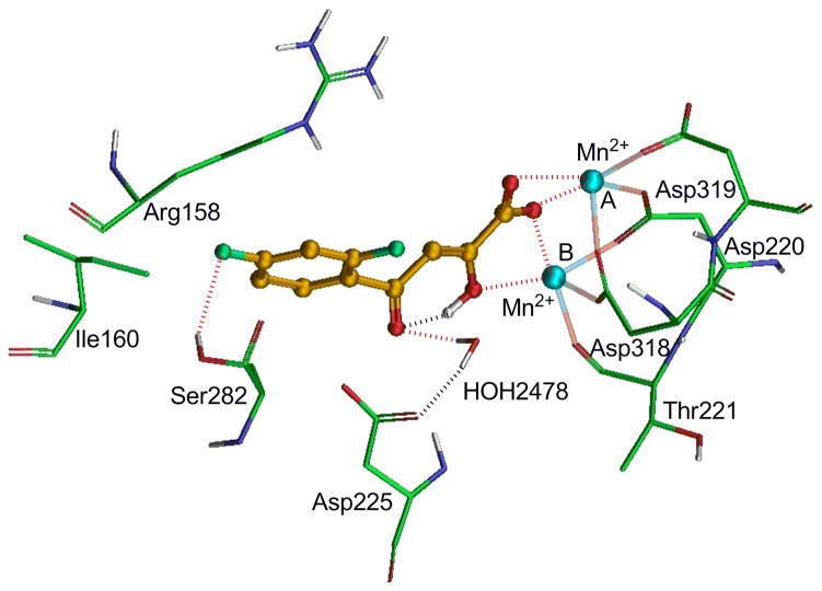
Figure 1.
Glide docking model of 24a within the active site of NS5B polymerase. Amino acid residues are shown in as stick model whereas inhibitor is shown as ball and stick model. The metal ions (Mn+2) A and B are shown in cyan color. Dotted black line indicates hydrogen bonding interaction whereas dotted red line indicates electrostatic interactions.

