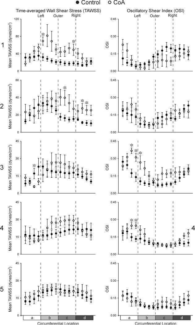
Figure 7.
Ensemble-averaged circumferential TAWSS (left column) and OSI (right column) plots comparing the six control subjects (solid circles) and age- and gender-matched coarctation patients treated by resection with end-to-end anastomosis (hollow circles) 1, 2, 3, 4, and 5 diameters distal to the LSA. a = anatomic inner left curvature, b = anatomic outer left curvature, c = anatomic outer right curvature, d = anatomic inner right curvature. * Statistically different from control subjects (P < 0.05). Data are expressed as mean ± SEM. Note the difference in scale for TAWSS 1 and 2 diameters distal to the LSA.

