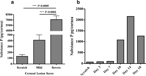
Figure 3.
Eyes with severe HSK lesions exhibit a higher level of SP in the cornea during the clinical phase of HSK. The corneas were grouped on the basis of the opacity and angiogenesis into mild and severe HSK on Day 15 postinfection. (a) SP levels in the corneal lysates grouped as mild or severe HSK were detected via ELISA. For mild HSK two corneas were pooled to obtain one sample whereas three corneas with severe HSK lesions were pooled to obtain one sample. A total of four to eight samples were obtained from two independent experiments. Results are reported as mean ± SEM. (b) Kinetics of SP amount in naïve and HSV-1–infected corneas were determined at different days postinfection. Six corneas were collected and pooled after infection with HSV-1 at the indicated time points. Graph represents the mean of two independent experiments.

