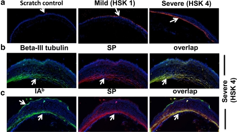
Figure 7.
Eyes with severe HSK lesions exhibit SP in the corneal stroma and colocalize with β-III tubulin and IA-b+ cell types. (a) Fifteen-micrometer frozen sections obtained from uninfected or the eyes with mild or severe HSK lesions were stained for SP (red) followed by acquisition on a confocal microscope (acquired at magnification ×10). (b, c) Fifteen-micrometer sections obtained from eyes with severe HSK lesions (corneal opacity score 4) on day 15 postinfection were stained with SP (red) and β-III tubulin (green) or IAb (green) obtained on Day 15 postinfection. Images were acquired on a confocal microscope (C1 Nikon; acquired at magnification ×20). Yellow: overlap between SP and β-III tubulin or SP and IAb staining.

