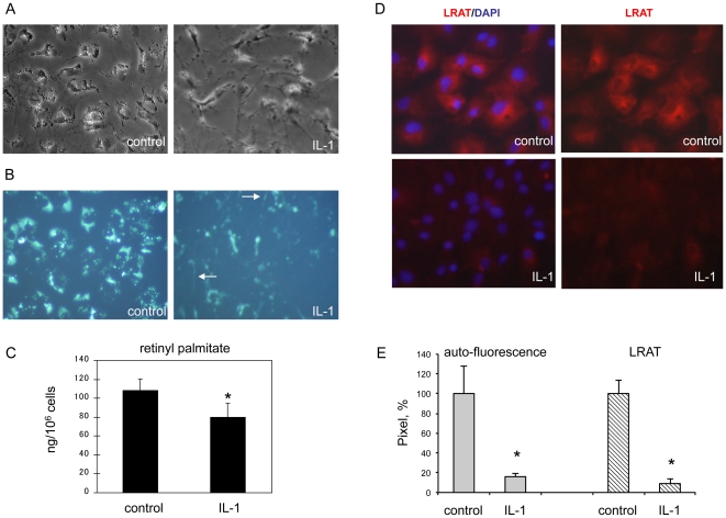
Figure 2. Interleukin-1 induced mobilization of retinyl ester in primary rat hepatic stellate cells is related to down-regulation of LRAT.
Primary rat HSCs were cultured on plastic plate and treated with or without IL-1α (10 ng/ml) for 3 days. (A) Phase contrast images of HSCs. (B) Vitamin A auto-fluorescence in HSCs under UV excitation. Arrows indicate the small droplets that undergo mobilization along cytoplasmic processes as visualized by UV excitation. (C) Retinyl palmitate storage was measured by LC/MS/MS analysis, by which all-trans retinoic acid was used as an internal control for sample normalization. Results are expressed as ng/106 cells. *P<0.05 (n = 6). (D) Immunofluorescence staining for LRAT (red) in HSCs. Nuclei are indicated by DAPI staining (blue). Original magnification ×200. (E) Densitometry (pixel) analysis of 5 independent images or fields. *P<0.05.

