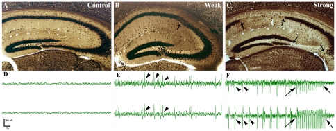
Figure 2. Anatomical and physiological changes in pilocarpine induced epilepsy.
Light micrographs of control (A) and epileptic (B, C) animals immunostained for NeuN. Animals survived for 1 month after pilocarpine treatment. Compared to control mice (A), in non-sclerotic “weak” animals (B) only mild, restricted changes (arrow) are seen occasionally in the hippocampus. In the sclerotic hippocampi of “strong” animals (C) a characteristic pattern of neuronal damage appears. The principal cell loss is over 50% (sclerosis) mostly in CA1 and CA3 (arrows) and in the hilus. In the EEG recordings of control animals no ictal or interictal acitivity can be seen (D), however, in weak animals (E) interictal spikes (arrowheads) occur frequently. In strong animals (F) recurrent seizures are observed in most cases, both interictal and ictal (arrows) activity appeares. An increase in frequency and decline in amplitude can be seen immediately preceding and after the seizures. Scale (A, B, C): 200 µm.

