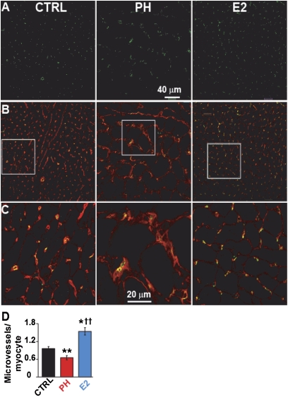
Figure 5.
Stimulation of cardiac neoangiogenesis by estrogen (E2). (A–C) Single confocal images of right ventricular sections of male rats immunostained for CD31 (green, A), overlay of CD31 and WGA (red, B) and at higher display magnification (C). (D) Quantification of microvessels/cardiomyocyte in control (CTRL; black bar), pulmonary hypertension (PH; red), and E2 (blue). *P < 0.05 versus CTRL, **P < 0.001 versus CTRL; ††P < 0.001 versus PH (n = 4 animals per group).

