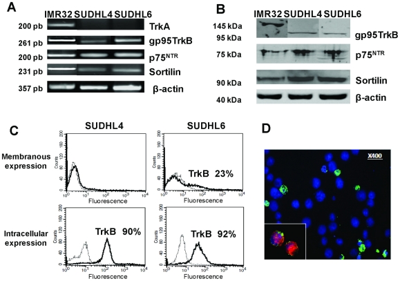
Figure 2. DLBCL cell lines express neurotrophin receptors.
A: RT-PCR detection of p75NTR, and its co-receptor sortilin, TrkA and truncated gp95TrkB mRNA was performed on SUDHL cells cultured with 10% FCS for 72 h. β-actin was included as a control of cDNA quality. B: The truncated TrkB, p75NTR and sortilin receptor protein expression was confirmed by western blotting of cell lysates. Blots were reprobed with anti- β-actin as a loading control. C: Flow cytometry analysis demonstrating TrkB membrane detection expressed in percentage of positive cells (bold line) in unpermeabilized SUDHL6 cells in contrast to SUDHL4 cells, whereas both cell lines expressed intracellular TrkB (dotted line: the isotypic controls). D: Immunofluorescence staining was performed, as described in Materials and Methods, in DLBCL cell lines that confirmed surface TrkB expression (in green) in some BDNF (inset in red) positive SUDHL6 cells (in blue: DAPI staining). The neuroblastoma cell line, IMR32, was used as a positive control. Data are representative of four independent experiments.

