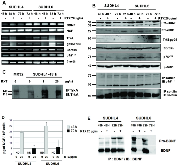
Figure 5. Effects of rituximab on neurotrophin production and Trk expression in DLBCL cells lines.
A: RT-PCR analysis of NGF, BDNF, TrkA, truncated TrkB, sortilin and p75NTR mRNA in SUDHL4 and SUDHL6 after 48 h and 72 h exposition to 20 µg/ml rituximab (RTX). β-actin was included as a control of cDNA quality. B: Western blott demonstrating in particular NGF protein expression following rituximab treatment and heterodimerization of p75NTR with sortilin in cell lysates. Blots were reprobed with anti- β-actin as a loading control. C: Enhanced TrkA expression induced by rituximab was observed for the less rituximab sensitive cell line, SUDHL4, after immunoprecipitation (IP) and immunoblotting (IB) with specific antibodies. D: NGF production was confirmed after rituximab exposure by detection with ELISA in cell supernatants of both cell lines, whereas it was undetectable (ND, non detected) in control cultures. Results are means ± SD of three independent experiments. E: BDNF secretion seemed to decrease in the most rituximab sensitive cell line, SUDHL6, as shown by western blott analysis of BDNF in cell lysate immunoprecipitates. Data are from one representative experiment out of four (RT-PCR, WB) or two (IP/WB) performed.

