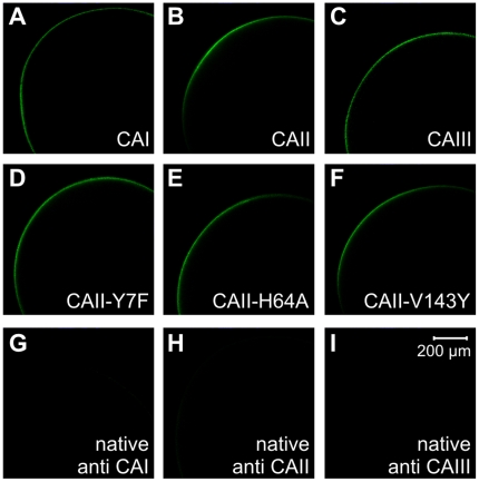
Figure 1. Fluorescent staining of CA isoforms and mutants.
Optical slices of oocytes, labeled via primary antibodies and Alexa Fluor 488-linked secondary antibodies against CAI (A), CAII (B), CAIII (C), CAII-Y7F (D), -H64A (E) and -V143Y-expressing oocytes (F). As control, a staining of native, uninjected oocytes or oocytes injected with H2O with primary antibodies against CAI (G), CAII (H) and CAIII (I) as well as corresponding secondary antibodies, respectively, was performed.

