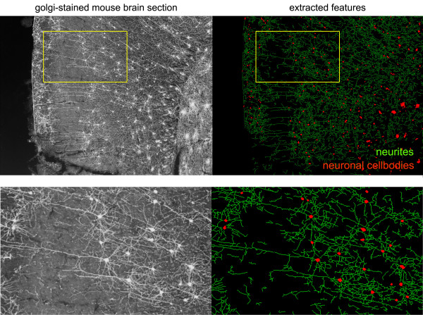
Figure 6.
Analysis of neuronal morphology in Golgi-stained mouse brain sections. Wild-type P26 mouse brains were stained using modified Golgi-Cox impregnation (FD NeuroTechnologies). NeuriteQuant is able to extract most features in images of silver-stained neurons that display a clearly defined soma and dendritic arbor (see enlarged region). During sectioning, neurites are often separated from their parent cell, therefore branching was not evaluated.

