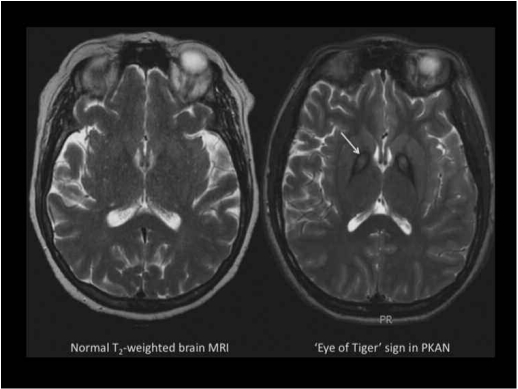
Figure 1.
(Left) Normal T2-weighted axial MRI. (Right) T2-weighted axial MRI from a patient with PKAN. In the globus pallidus, the region of central hyperintensity represents tissue edema and precedes the emergence of iron accumulation, which is seen as the surrounding hypointense region. This pattern is called the ‘eye of the tiger’ sign.

