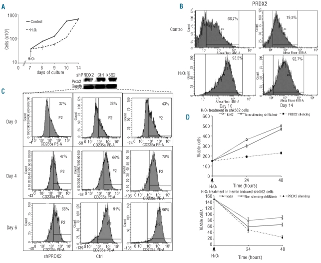
Figure 4.
Effects of oxidative stress on PRDX2 protein expression in normal erythroid precursors and in K562 cells with PRDX2 depletion. (A) Effect of H2O2 (16 μM) on cell proliferation of control erythroid precursors. Data are presented as mean ± SD (n=3). (B) Flow-cytometric analysis of PRDX2 in normal erythroid precursors exposed to H2O2 (16 μM). Each culture was divided into two separate cultures with and without H2O2 (16 μM). The ordinate represents the number of cells displaying the fluorescent intensity given by the abscissa (see also Online Supplementary Data for cell gating strategy). The figure shows the results from one typical erythroid culture representative of three studied. (C) Effect of PRDX2 depletion during erythroid differentiation of K562 cells. Human K562 cells were induced to differentiate with hemin (50 μM) after 24 h of transfection with K562 wild type and non-silencing vector as control (Crtl), and shRNAmir plasmid against PRDX2. Samples were collected at specific time points (after transfection): before hemin addition, and at days 4 and 6 after hemin addition. Erythroid differentiation was assessed by FACS analysis for glycophorin A (CD235A). Analyses were performed using a FACSCalibur (Becton Dickinson, San Jose, CA, USA) with CELL Quest software, version 3.3, after gating for viable cells. The upper panel shows the immunoblot analysis with specific anti-PRDX2 antibody in silenced cells (shPRDX2), non-silencing vector as control cells (Crtl) and K562 cells. One representative gel of three other with similar results is presented. (D) Effect of H2O2 on cell viability of parental (upper panel) and hemin-induced (lower panel) k562 cells after silencing. Data are presented as mean ± SD (n=3). Cells were treated by 30 min incubation with 50 μM H2O2 (
 K562 wild type, □Ctrl, non-silencing vector, and ■shPRDX2 after transfection; ▵K562 wild type,
 Ctrl, non-silencing vector, and ▴ shPRDX2, hemin-induced cells). Cell growth was evaluated by seeding the cells, after repeated washing, in Iscove’s complete medium (1.5x10⁵ cells/mL); the number of viable cells was evaluated at 24 and 48 h by the trypan blue dye exclusion test;47 Data are presented as ratio of trypan blue-positive cells; n=3 for all points.

