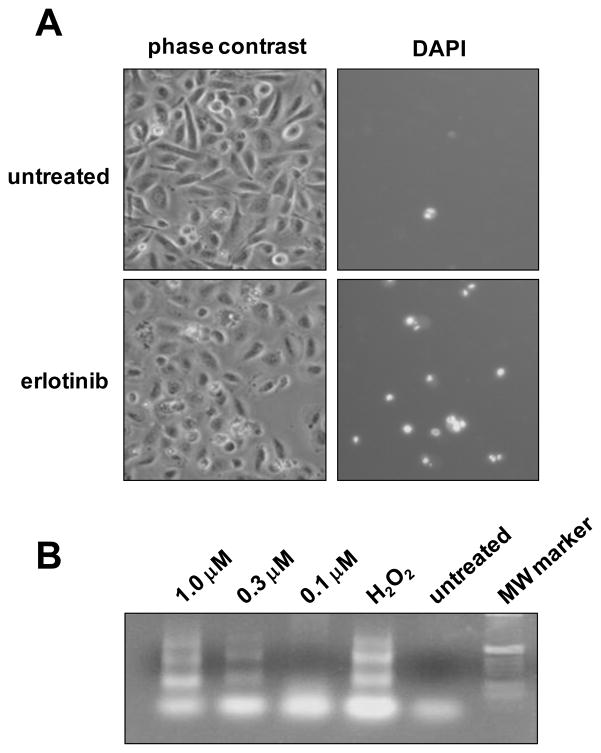
Figure 2.
HPV-16 E6/E7 expression sensitizes cervical cells to erlotinib-mediated apoptosis. A. Phase contrast micrographs (left) and fluorescent micrographs (right) of E6/E7-infected cells that were untreated (top) or treated with 1.0 μM erlotinib for 48 hours (bottom) and then stained by DAPI to label cells undergoing apoptosis. B. TUNEL assay of cells treated with erlotinib. Cells infected with HPV-16 E6/E7 were treated with EGFR inhibitor for 24 hours and analyzed for apoptosis using a DNA Ladder Assay PCR kit to detect inter nucleosomal DNA fragmentation. H2O2 served as a positive control for apoptosis.

