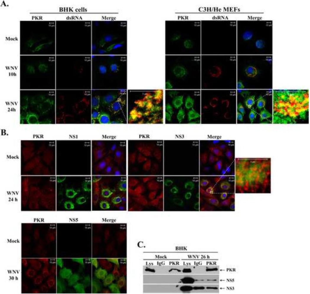
Figure 2. PKR colocalization with sites of WNV replication.
(A) Analysis of PKR colocalization with viral replication complexes. BHK cells and MEFs were mock-infected or infected with WNV Eg101 (MOI of 5). At 10 and 24 h after infection, cells were permeabilized, fixed, and blocked overnight. Cells were stained with anti-PKR and anti-dsRNA antibodies and then AlexaFluor488 (green) and AlexaFluor594 (red) conjugated secondary antibodies, respectively. Cell nuclei were stained with Hoechst. (B) Analysis of PKR colocalization with viral nonstructural proteins. BHK cells and MEFs were mock-infected or infected with WNV Eg101 (MOI of 5). At 24 and 30 h after infection, cells were permeabilized, fixed, and blocked overnight. Cells were stained with anti-PKR and either anti-NS5, anti-NS3 or anti-NS1 antibodies and then AlexaFluor488 (green) and AlexaFluor594 (red) conjugated secondary antibodies, respectively. Cell Nuclei were stained with Hoechst. (C) Analysis of PKR interaction with viral NS3 or NS5 proteins in infected cells. BHK cells were mock-infected (M), or infected with WNV Eg101 (MOI of 5). At 26 h after infection, cells were lysed, S2 fractions were prepared and rabbit anti-PKR antibody or a rabbit IgG was used for immunoprecipitation. Immunoprecipitated proteins were separated by 10% SDS-PAGE and detected by Western blotting using anti-NS5 or anti-NS3 antibodies.

