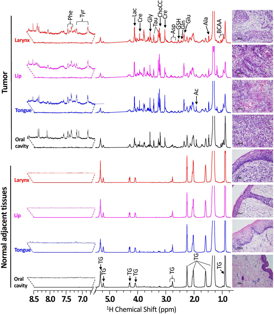
Figure 1.
Representative 600 MHz 1H HR-MAS CPMG NMR spectra (area normalized) of matched NAT and HNSCC tissues from various anatomical sites of head and neck involving oral cavity, tongue, lip and larynx of four different patients. The vertical scales of all the spectra were kept the same and the chemical shift region from 5.2 – 6.5 ppm (spinning side bands) is not shown in all the spectra. The intensity of peak in the chemical shift region 6.7 – 8.5 ppm was increased equally in all spectra to show the low-abundant taurine and phenylalanine. The triglyceride signals were indicated as ‘TG’. The corresponding H&E photomicrographs of post HR-MAS NMR tissues are shown alongside of each 1H CPMG spectrum. All the NAT are composed of parakeratinized stratified squamous epithelium and underlying fibrovascular connective tissue. However, tumor tissues were composed of squamous cell carcinomas with neoplastic cells demonstrating varying degrees of epithelial differentiation invade the adjacent connective tissue predominantly as nests, sheets or single cells.

