
Fig 1. Influence of topographic surfaces on cell morphology.
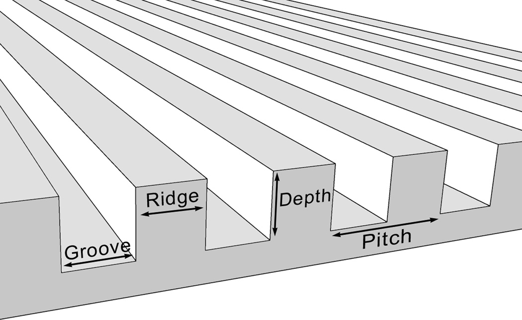
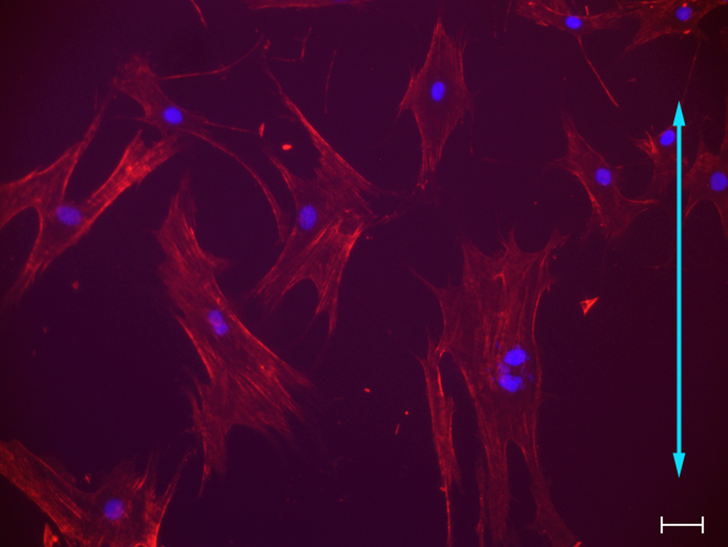
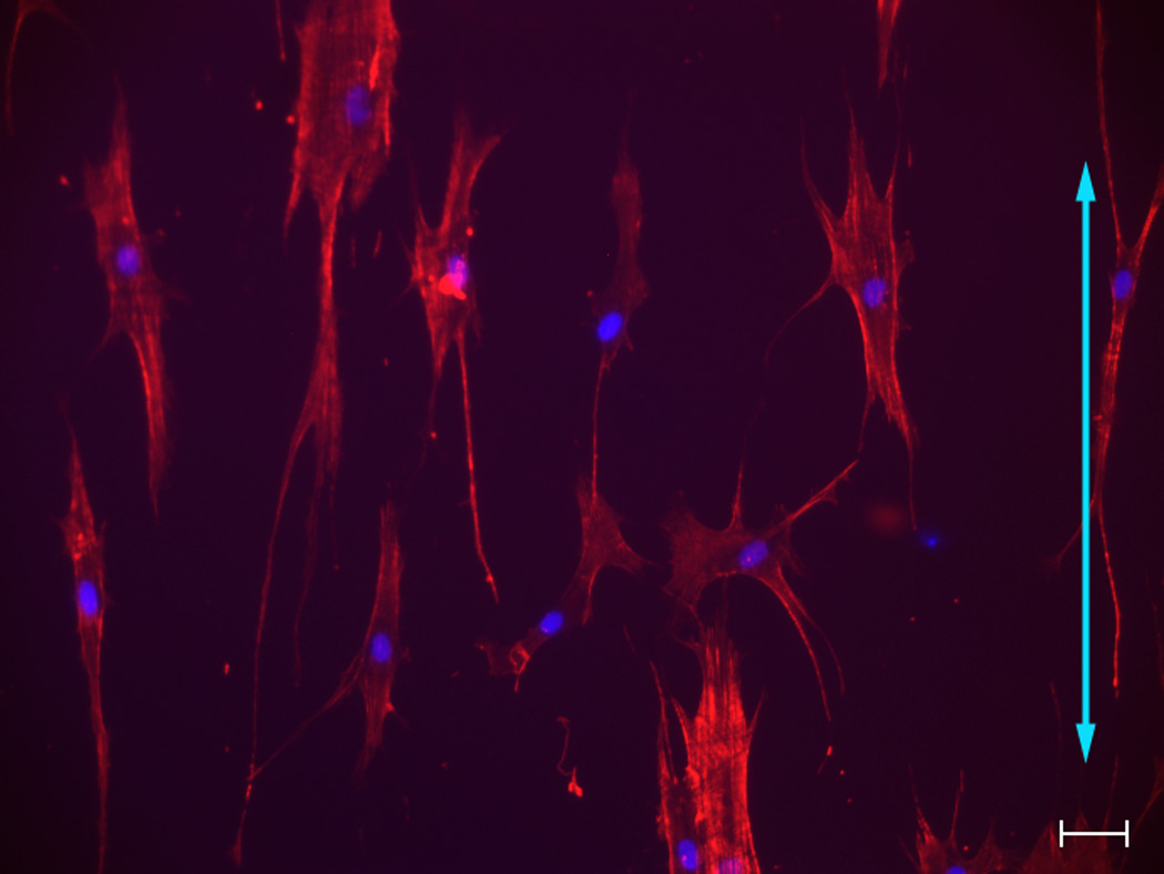
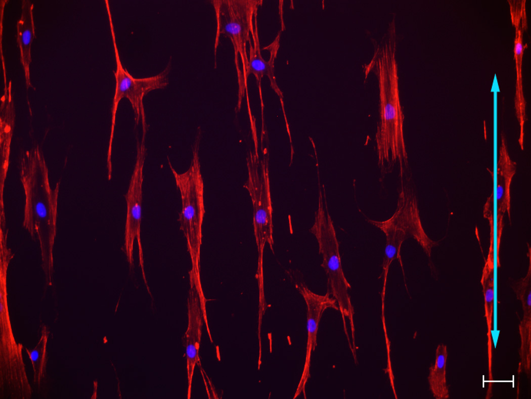
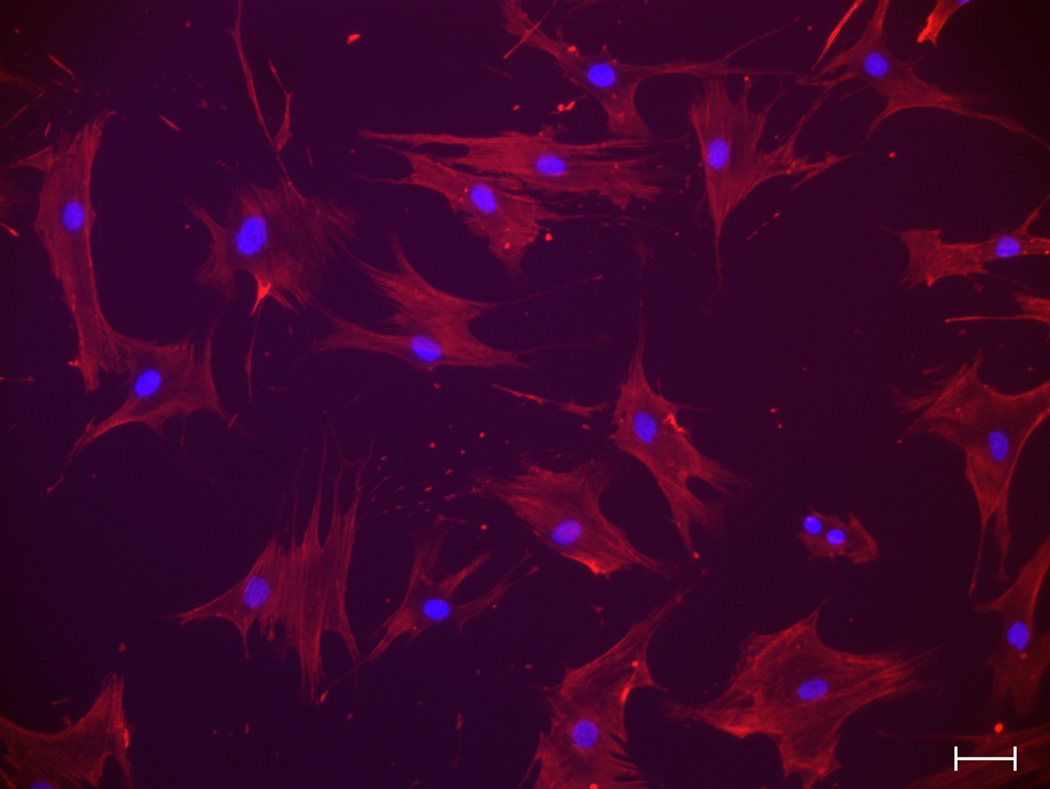
Pitch is defined as the sum of the width of a ridge and a groove as shown on the schematic diagram of a topographic surface (A). The depth for all the topographic surfaces was 300 nm. Representative images of hMSCs were taken with a Zeiss Axiovert 200 M inverted microscope on 400 nm pitch (B), 1400 nm pitch (C), 4000 nm pitch (D) and planar control (E). hMSCs on 400 nm and planar surfaces showed a more rounded and flattened morphology while hMSCs on 1400 and 4000 nm pitch showed an elongated morphology. Each picture was taken at 10 X magnification. Double-headed arrows represent direction of the underlying topography of ridges and grooves. Scale bar: 50 µm.