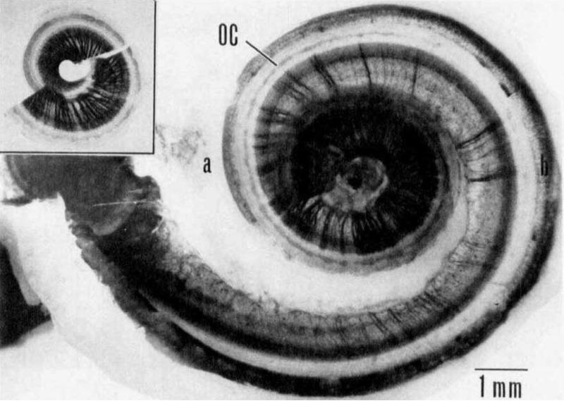
Fig. 1.
Example of patchy nerve survival in an ototoxically deafened human cochlea as seen in a surface preparation by Johnsson and colleagues (1981). The inset shows the upper half of middle and apical turns of the cochlea. Degeneration is more severe in the basal turns (main figure). Nerve fibers in the osseous spiral lamina are stained with osmium tetroxide. OC = supporting cells in the Organ of Corti. No hair cells were found. Reproduced, with permission, from Acta Oto-Laryngologica, 1981, Supplement 383, page 12, Figure 9.

