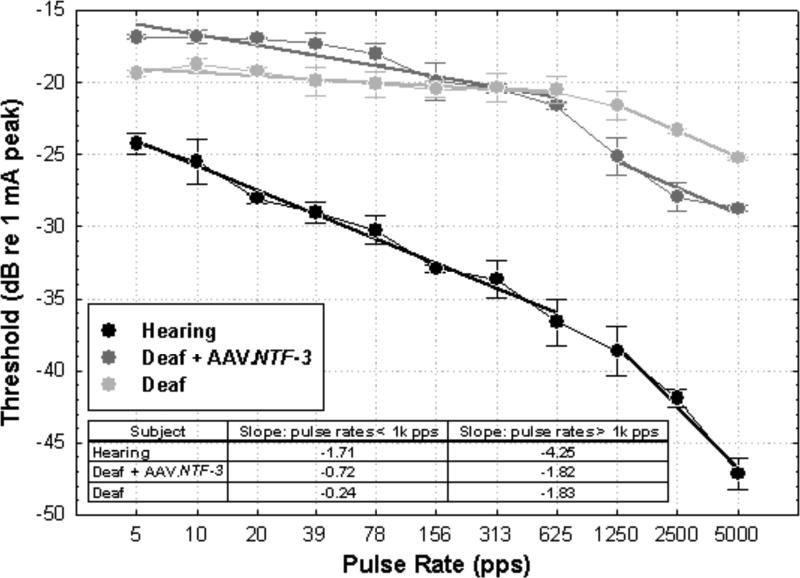
Fig. 6.
Psychophysical detection thresholds for monopolar pulsatile stimulation of Electrode B as a function of pulse rate for the three animals for which histology is shown in Fig. 3. Stimuli were 25 μs/phase biphasic pulses delivered by a controlled-current stimulator (built in house). Stimulus levels in μA were controlled in 1 dB steps. Detection thresholds (50% correct detections) are presented in dB relative to 1 mA peak. Pulse-train duration was 200 ms. Means and standard deviations for three repeated measures are shown. Other details of the psychophysical training and testing procedures are given in Kang et al., 2010. Best-fit linear regression lines were fit to the data points below 1000 pps and separate lines were fit to the data above 1000 pps. These slopes, in dB per doubling of pulse rate, are given in the inset table. Differences between the deaf and hearing animals in the slopes and levels of these functions are similar to those reported in Kang et al., 2010. Experiments are currently in progress for two other guinea pigs that were deafened and inoculated with AAV.NTF-3. Slopes for these animals for pulse rates below 1 kpps were similar to those for the deafened AAV.NTF-3 treated animal in this graph (-0.88 and -1.36 dB/doubling).

