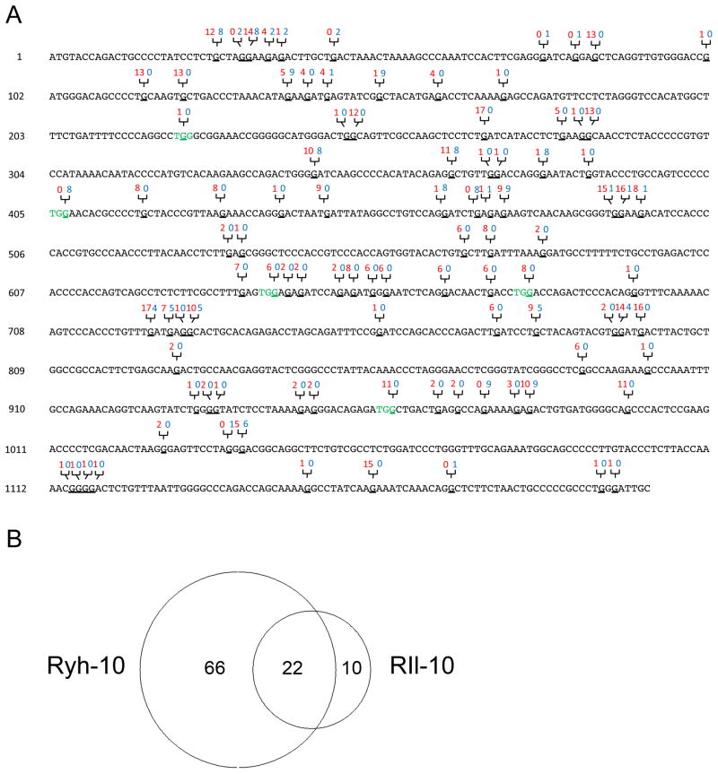
Figure 3. Locations and numbers of A3-mediated G to A mutations in a 1.2 kb region of XMRV pol sequence.
(A) Nucleotide locations of G to A mutations in the 1.2 kb XMRV pol proviral sequence from both rhesus macaques. The number of clones from RIl-10 (red) and RYh-10 (blue) that contains the specific mutation are labeled above the sequence. Codons mutated by A3 to stop codons are shown in green. Mutated G nucleotides are underlined. (B) Shared and unique numbers of G to A mutations from both rhesus macaques depicted in a Venn diagram generated online at the website http://jura.wi.mit.edu/bioc/tools/venn.php (Whitehead Institute for Biomedical Research, Cambridge, MA).

