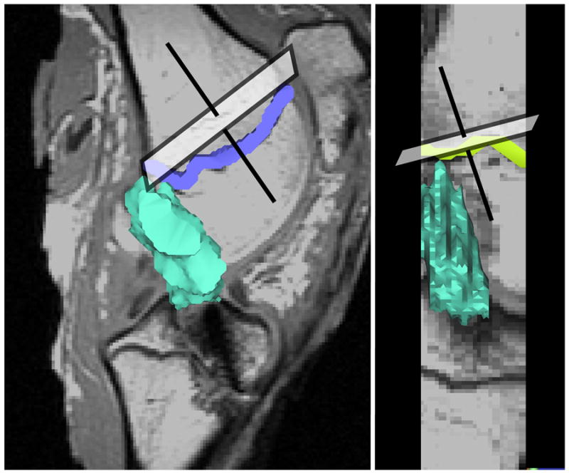
Figure 1.

Sagittal oblique images were oriented perpendicular to the distal femoral epiphyseal plate in both the coronal and sagittal planes. The intra-articular volume of the ACL graft (shown in green) was determined by segmenting the ACL within each slice and stacking the slices.
