Figure 2.

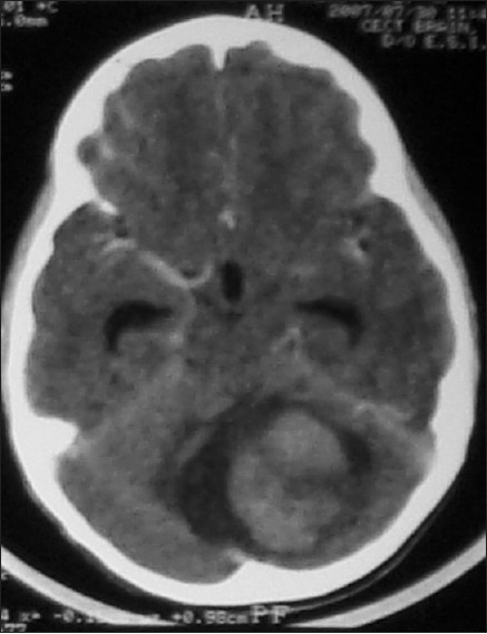
CT scan showing a large solid enhancing cerebellar lesion with areas of hypodensity within. There is a small cystic area around the lesion. At surgery, the solid tumor was radically excised. Histologically, it was a fibrillary astrocytoma

CT scan showing a large solid enhancing cerebellar lesion with areas of hypodensity within. There is a small cystic area around the lesion. At surgery, the solid tumor was radically excised. Histologically, it was a fibrillary astrocytoma