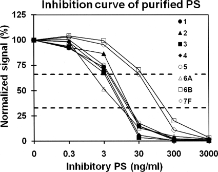
Fig. 2.
Normalized fluorescence signals of the PS-coated latex particles in the presence of varying concentrations of homologous free inhibitory PS in solution. The serotypes of inhibitory PS are given in the key. Two horizontal dashed lines indicate 33% and 67% of normalized signals, the values that were used as cutoffs.

