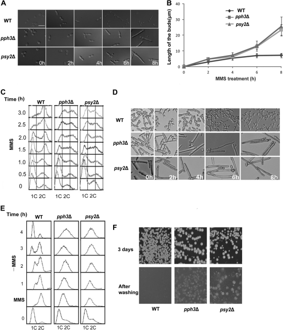
Fig. 4.
pph3Δ and psy2Δ cells exhibit irreversible pseudohyphal growth and cell cycle arrest when treated with MMS. (A) WT (SC5314 or BWP17), pph3Δ (SJL3), and psy2Δ (SJL6) yeast cells were grown in YPD medium supplemented with 0.02% MMS at 30°C. Cells were collected for microscopic examination at timed intervals. Bar = 5 μm. (B) Comparison of filament lengths of cells treated with MMS. Filament length was measured using ImageJ (http://rsbweb.nih.gov/ij/index.html). The data points show the average of 30 cells. The experiment was repeated 3 times. The error bars indicate 1 SD. (C) Cells were treated as described for panel A and harvested at intervals for flow cytometry analysis. (D and E) The same cells used in panel A were incubated in YPD medium containing 0.02% MMS for 6 h before washing the cells with fresh YPD and inoculating the MMS-treated cells into MMS-free YPD medium. Aliquots were harvested at intervals for microscopy and flow cytometry analysis. (F) pph3Δ and psy2Δ cells invade agar during recovery from DNA damage. BWP17 and mutant (SJL3 and SJL6) cells that had been grown in YPD medium containing 0.02% MMS for 6 h were spread onto YPD agar plates and incubated at 30°C. As ∼90% of the mutant cells lost viability, after the MMS treatment, the cells were first concentrated by ∼10-fold before being spread onto the plates so that more colonies would grow. The plates were photographed after 3 days and then washed to remove cells on the agar surface and rephotographed.

