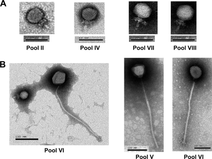
Fig. 1.
Transmission electron micrographs of virulent bacteriophages against S. epidermidis ATCC 35983 isolated from the anterior nares of humans. Panels: A, Podoviridae; B, Siphoviridae. Roman numerals represent pooled samples (see Table 1 and text). Two pictures from pool VI are shown in panel B, one of a Siphoviridae phage alone and the other of a Siphoviridae and a Podoviridae phage.

