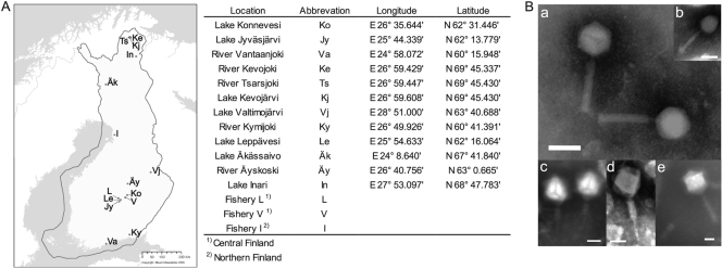
Fig. 1.
(A) Sites in Finland where flavobacteria and their phages were isolated. Sampling sites are marked on the free map obtained from the National Land Survey of Finland (Maanmittauslaitos, 2005). The sites and their abbreviations and coordinates are listed on the right. (B) Electron micrographs of purified and negatively stained tailed Flavobacterium phages. (a) FCV-1; (b) FCL-2; (c) FJy-3; (d) FKo-2; (e) FKy-1. Bar, 50 nm.

