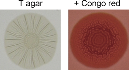
Fig. 5.
Colony morphology of an E. coli isolate displaying the rdar morphotype. Colony morphology of an E. coli rdar+ isolate is shown after growth at 28°C for 72 h on tryptone agar or tryptone agar supplemented with Congo red. Note the distinctive, patterned appearance of the colony and deep red associated with extracellular matrix production and formation of the rdar morphotype.

