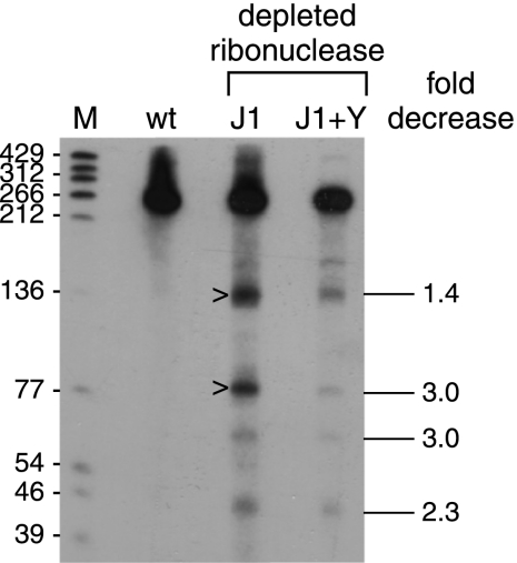
Fig. 2.
Analysis of ΔermC mRNA decay fragments probed with a 36-nt 3′-terminal probe that was complementary to ΔermC mRNA nt 205 to 240. The strain types are indicated above each lane: wt, wild type; J1, RNase J1 limited; J1+Y, RNase J1 and RNase Y limited. Marker lane M contained 5′-end-labeled TaqI fragments of plasmid pSE420 DNA (2), with the sizes of these fragments indicated on the left. On the right is the fold decrease of each of four prominent decay intermediates in the J1+Y lane relative to the J1 lane and normalized to the amount of full-length RNA (average of results from four experiments).

