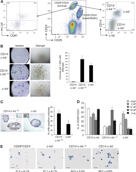
Fig. 1.
Isolation of a distinct alveolar progenitor cell using the CD14 and c-kit markers. (A) Representative FACS dot plot showing the expression of CD29 and CD24 in Lin− (CD31− CD45− TER119−) cells isolated from 8-week-old virgin mouse mammary glands (middle panel). Representative FACS dot plots showing CD61 and c-kit expression (left panel) or CD14 and c-kit expression (right panel) in Lin− CD29lo CD24+ cells in mammary glands taken from an 8-week-old virgin mouse. (B) Colony-forming capacity of c-kit/CD14 subpopulations plated on fibroblast feeder layers or embedded in Matrigel, as indicated. Right panel, histogram showing the colony formation capacity of c-kit/CD14 cells plated in Matrigel. Data represent the means ± standard error of the means (SEM) of results of three independent experiments. (C) Representative anti-milk protein immunostaining of CD14+ c-kit−/lo and c-kit+ colonies grown in Matrigel for 14 days. Left inset shows staining for the isotype control antibody; right inset shows milk staining of a section from an 18.5-day-pregnant-mouse mammary gland. Scale bars, 60 μm. Right panel, bar chart showing the percentage of milk protein-positive colonies observed 14 days after plating of CD14+ c-kit−/lo and c-kit+ cells in Matrigel (means ± SEM of results of three independent experiments). (D) Histogram showing the percentage of c-kit/CD14 cells in the Lin− CD29lo CD24+ population at different stages of development: virgin (V), pregnancy (dP), and lactation (dL). Results represent the means ± SEM of results for three to five animals per group. (E) β-Galactosidase activity in freshly cytospun CD29hi CD24+ (MaSC-enriched), c-kit+, CD14+ c-kit−/lo, and CD14− c-kit− cells from Gata-3+/nlslacZ mice. Scale bars, 25 μm.

